Cervical Anatomy
The cervical spine includes the first seven vertebrae. Located between the head and the relatively stiff thoracic spine, the cervical spine can be subject to both wear and tear problems (degenerative) as well as trauma (injury).
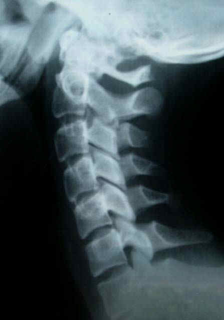
On this normal xray, to the left, you will note that the disc spaces are all about the same thickness at each level, and there is a gentle curve, hollow to the back of the neck.
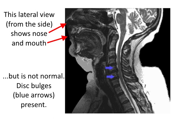
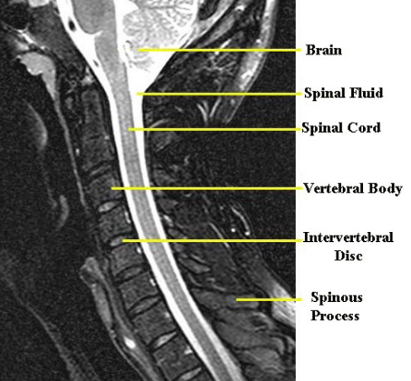
Important features of the cervical spine are shown on these MRI images with an example that shows a broader view (showing face, chin, and tongue, and also some disc bulges) below right, and with normal below left,

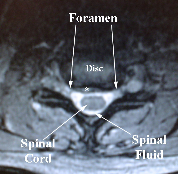
This MRI image is from a cross section of the neck, as if there was a cut with a guillotine.
The orientation of the picture is that the front is towards the top, and the back is towards the bottom of the image.
Note that the disc and the vertebral bodies are in the center of the neck.
This illustration below shows how the cuts through the spine correlate with the images.
Like slicing a salami, the cuts go either through the foramen, the opening through which nerve exits (here with this type of section marked “B”), or through a part of the bone called the pedicle (like the slice labeled “A.”)

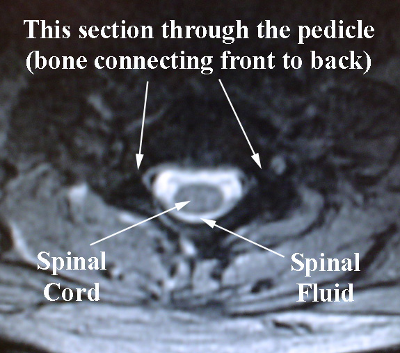
Cut through the pedicle: Type A slice
Here is an example of a type “A” slice, with bone connecting the vertebral body in front, with the back part of the spine called the lamina.
Cut through the foramen: Type B slice
Here is an example of a type “B” slice which cuts through the foramen (the opening through which nerve exits).