Foraminotomy (opening up the foramen)
The foramen is the hole or opening through which the nerve root passes as it leaves the spinal canal. Sometimes this opening can become more narrow, either from a bulging disc in the front, or from an arthritic facet joint in the back.
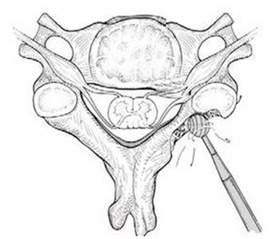
The term “foraminotomy”, which refers to a procedure usually done from the back side of the patient, involves removal of some bone and soft tissue from the back of this opening, relieving pressure on the exiting nerve root.
In this spinal model, the superior facet, which is the facet that goes up towards the head, can become enlarged due to an arthritic process. Another source of potential compression on the nerve might be from the disc (white cushion between the bones here) bulging in a backward direction.
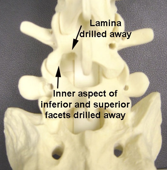
In the foraminotomy procedure, some of the bone is drilled away from the lower edge of the lamina (the bone on the back of the spinal nerves) and from the inner edge of the facets, both the inferior (the one that goes downward) and the superior (the one that extends upwards) facet.
In the foraminotomy procedure, as shown here from a dorsal (back side) view, some of the bone is drilled away from the lower edge of the lamina (the bone on the back of the spinal nerves) and from the inner edge of the facets, both the inferior (the one that goes downward) and the superior (the one that extends upwards) facet.
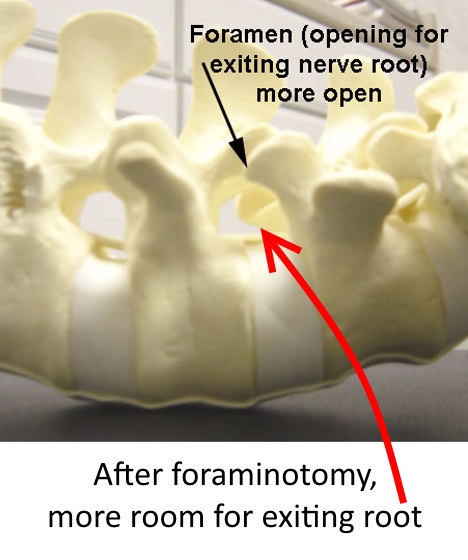
The net effect of this bone removal is that the opening is larger for the nerve root to pass through. The foraminotomy procedure has been done on this spine model as indicated by the arrows.
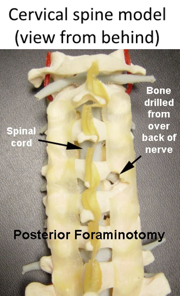
In this model of a cervical spine, viewed from behind, the spinal cord and nerves are colored gray.
For the segment where the path for the exiting nerve root is too tight, the foraminotomy will make the opening through which the nerve exits bigger by drilling bone off the back side of the exiting nerve root, thus relieving any pressure on the nerve root.