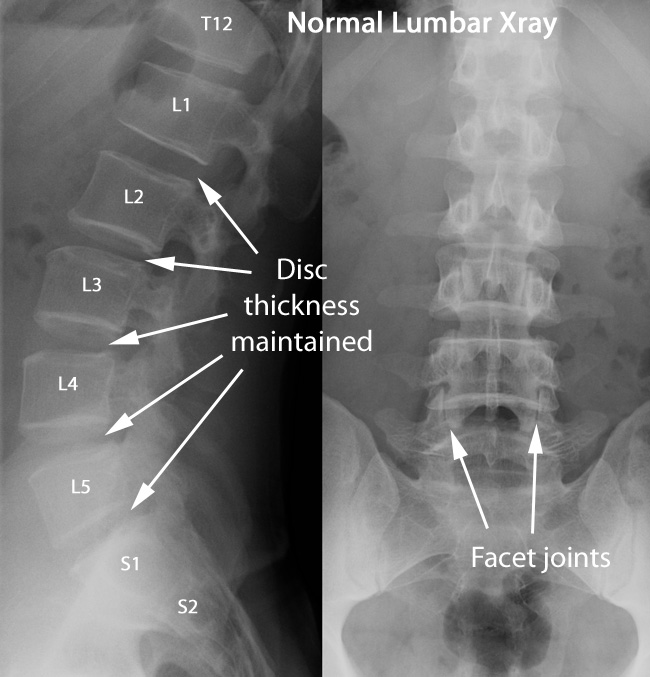
These X-rays to the right show normal findings. On the side view (also called a “lateral” view), the spaces between the vertebral bodies appear dark because the disc material doesn’t have calcium, and the X-ray beams go through them. If the discs were degenerated, the spaces would be thinner, and the bones would appear closer together.
Additionally, the spine has a curve on this view that is hollow to the back, bowed out towards the front. This curve is called "lordosis."
On the other film to the far right, which is a front view (called “AP” view which stands for Anterior Posterior, the direction of the X-ray beam), note that the facet joints can be seen. Also, there is no curvature of the spine to one side or the other, which if present, is called "scoliosis."
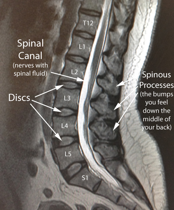
This picture, which is from an MRI on a patient with normal anatomy, is taken with a cut right down the middle, dividing equally in half between the right and the left. Images made by slicing in half the right and left are called sagittal images.
For the discs, which have a structure like a jelly donut, the center (jelly) part can be seen as more white, with higher water content, as different than the rim around it (the dough) which looks darker. The discs are contained in the area between the bones, and no bulging or degeneration is seen here.
The spinal canal is the place where all the nerves continue after the end of the spinal cord, which ends about the L1 vertebra.
The white around the nerves is due to the spinal fluid.
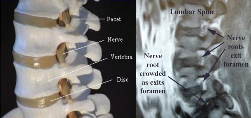
If the sagittal (those cutting the body in half, separating right and left) images are made a little to one side or the other, they can help to give imaging details of the foramen, which are the holes in the spine through which the individual nerve roots exit.
In the picture showing the spine model (below on the left), the nerve root can be seen coming out behind the disc and in front of the facet joint. In the MRI image (below on the right), you can see three foramen with adequate opening for the exiting nerve roots (the darker spots at the tips of the arrows, surrounded by white fat), but for the lowest foramen on this image, the nerve root is crowded and compressed (a condition known as stenosis). The patient would most likely feel pain and possibly have muscle weakness in the area supplied by that particular nerve root.

This view is taken in cross section separating the top and bottom of the patient, as if being sliced like a salami, and this view is what one of the slices would look like.
The facet joints, which are joints that have cartilage surfaces, like your finger or knee, have a small amount of joint fluid. Since this joint fluid, along with the joint cartilage, have higher water content than bone, the MRI has an appearance of more white color between the two sides of the facet bones, with the bone appearing more black.
To see information about abnormal facets, click here.
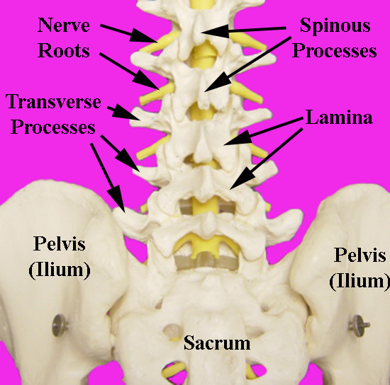
Various aspects of lumbar anatomy are illustrated here with these models.
The facet joints are joints on the right and left of the back of the spine, each about a half inch off the midline, and these joints slide apart or overlap more when the spine either flexes forward, or bends backward, respectively.
The spinous processes are the bumps you feel down the back of your spine in the midline.
The lamina is the sheet of bone covering the spinal nerves from behind.
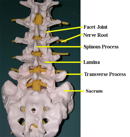
This picture of another spinal model shows the same parts.
The transverse processes are a part of the lumbar spine bones that are like miniature ribs, and often in spinal fusion operations, one of the goals is to get bone to grow between these parts of the vertebra.