Microdiscectomy, Minimally Invasive
“Discectomy” refers to removing disc. This procedure is usually done when a piece of disc is bulging out or has broken off and is touching or compressing a nerve.
"Micro" refers to the fact that usually the microscope is used for magnification, and that only a small incision in most cases is required.
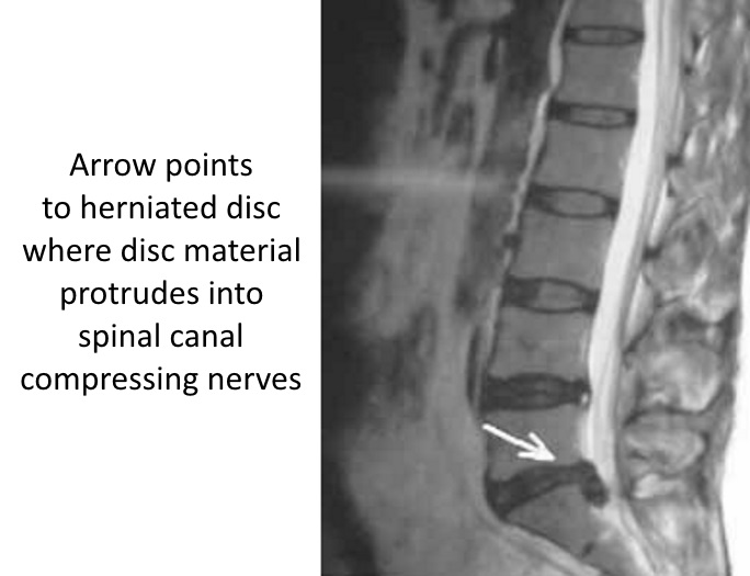
In the case shown here, the MRI shows the disc material extruded from the L5S1 disc.
This condition can cause back pain, but nerve compression usually causes pain down the leg, often referred to as "sciatica."
This type of surgery does not involve fusing the spine.
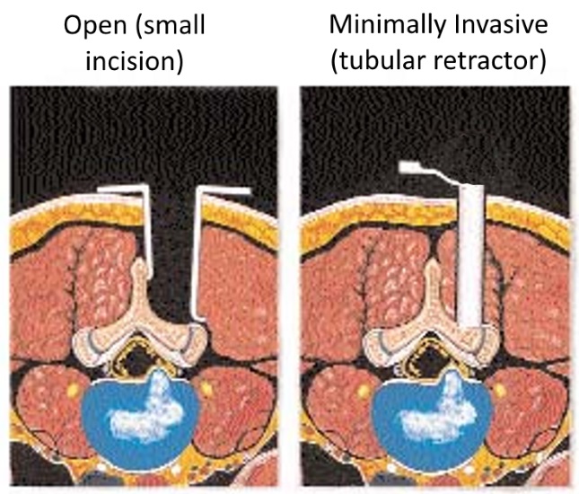
By using a minimally invasive approach, the dissection of the covering soft tissues can be minimized, leaving more blood supply to the surrounding tissues often associated with less pain and a quicker recovery, since less tissue is disrupted in the exposure.
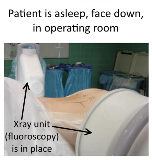
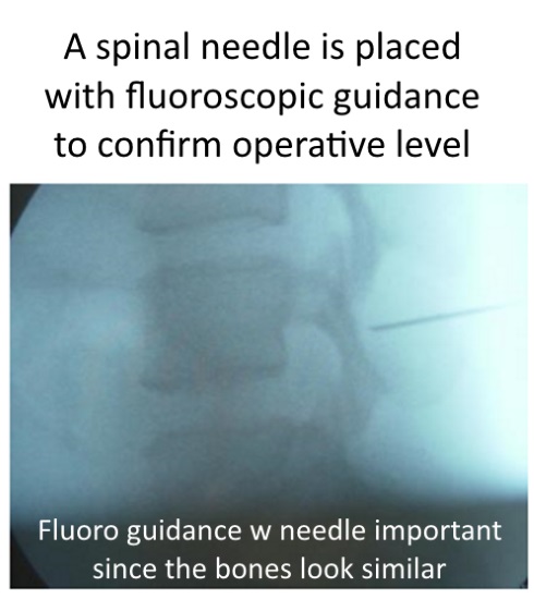
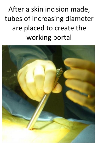
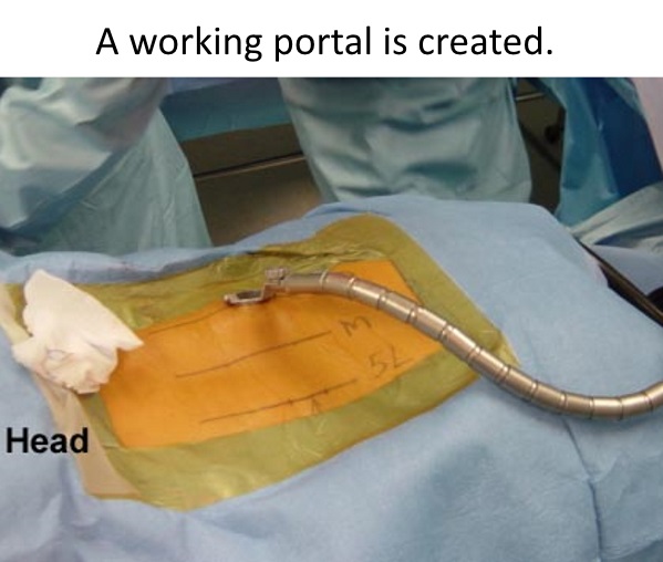
Technique notes: Minimally Invasive Microdiscectomy
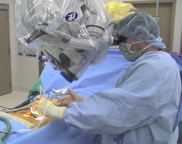
Operating microscope is brought in.
Procedure details
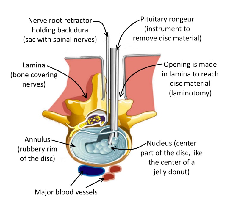
This diagram shows the important details of the surgery. After gaining exposure to the posterior (back side) of the spine, an opening is made in the posterior bone (lamina) called a laminotomy. The dura, which is the sac of nerves, is held towards the midline by a retractor, and the tear in the annulus (the dough of the jelly donut) can usually be seen. The disc material is removed with various instruments, like the one shown here, a small grabber called a pituitary rongeur.
While some surgeons take out only the bulging piece, I tend to remove all the nucleus to have lower risk of recurrent herniation.
By removing the compression on the involved nerves, the patient will usually wake up in the recovery room with their leg pain being gone.