Spine Anatomy
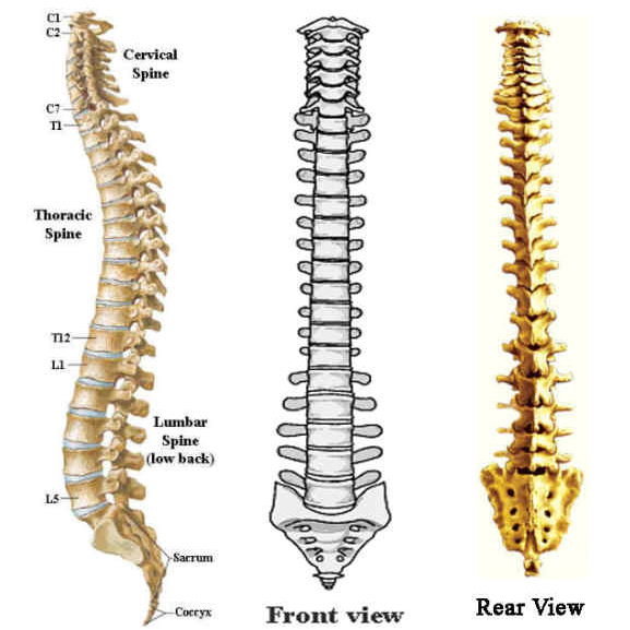
The spinal column, which is a series of bones that supports the body, maintains posture, and protects the spinal cord and nerves, is divided into several main sections:
- Cervical (neck)
- Thoracic (chest)
- Lumbar (low back)
- Sacrum (base of the spine, incorporated into the pelvis)
- Coccyx (tailbone)
For video from another site that has animations showing details of spinal anatomy, click here.
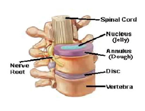
The disc, which is a cushion between the vertebral bodies, has a structure like a jelly donut, with a rubbery rim (annulus fibrosus) and a jelly center (nucleus pulposus). The center has higher water content and in a dissection would look like crab meat.
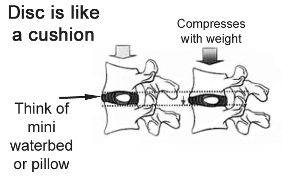
Another way to think of the disc would be like a little water bed: it can move and accommodate changes of position, and if the water dried up, the height of the disc would be less (like a flat tire).
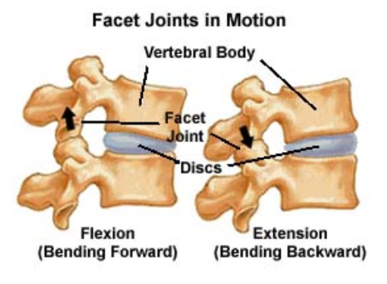
In the back part of the spine, there are joints that are in contact with the corresponding joints of the vertebra above and below. These joints are called the facet joints. These joints are covered with the type of cartilage like on the end of a chicken bone. This joint surface, which is normally smooth, can with wear and tear, become arthritic and rough, and be a source of pain.
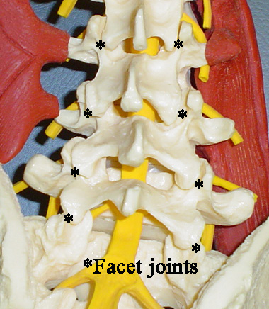
FACET JOINTS
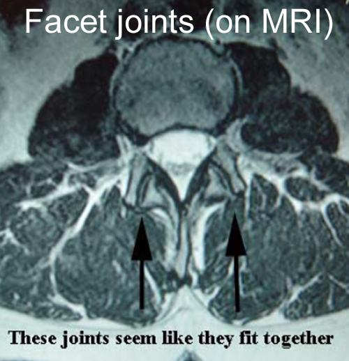
The best way to three dimensionally evaluate facet joints is with a cross sectional view, as shown here with an MRI scan. CT can also be helpful. Here, the two parts of the facet joint seem to fit together
To see worn out facets, click here.
FORAMEN
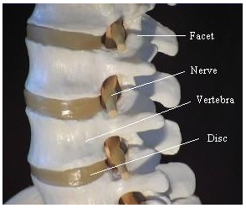
The opening in the spine through which each nerve root exits the spinal canal is called the foramen. The front of the foramen is the vertebral body (here marked “Vertebra”). The intervertebral discs are also within the foramen, so any bulging of a disc in this area could contact or compress the nerve root. Behind the foramen is the facet joint.
To see about narrowed formen, click here.
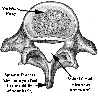
Behind the vertebral body is the place where the spinal cord and nerves reside, an area called the spinal canal.
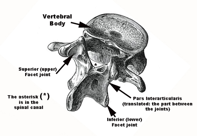
For each spine bone (vertebra), there is a facet that goes upwards (the superior facet) and one that goes downward (the inferior facet). The part in between these facet joints, which is an important landmark, is called the pars interarticularis.
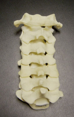
The bones in the neck have a somewhat different shape, but the same components: vertebral bodies separated by discs in front, facets in back, nerve roots leaving at every level.
For a video on cervical spine motion,here
For a more expansive explanation from the website
www.allaboutbackandneckpain.com, please here